 We learned a ton of stuff about the eye on today’s Healthy Matters broadcast. We mostly focused on glaucoma (good pun, eh. . . see how I said focused while talking about vision . . . oh never mind). We also touched on macular degeneration, cataract, and other topics raised by listeners.
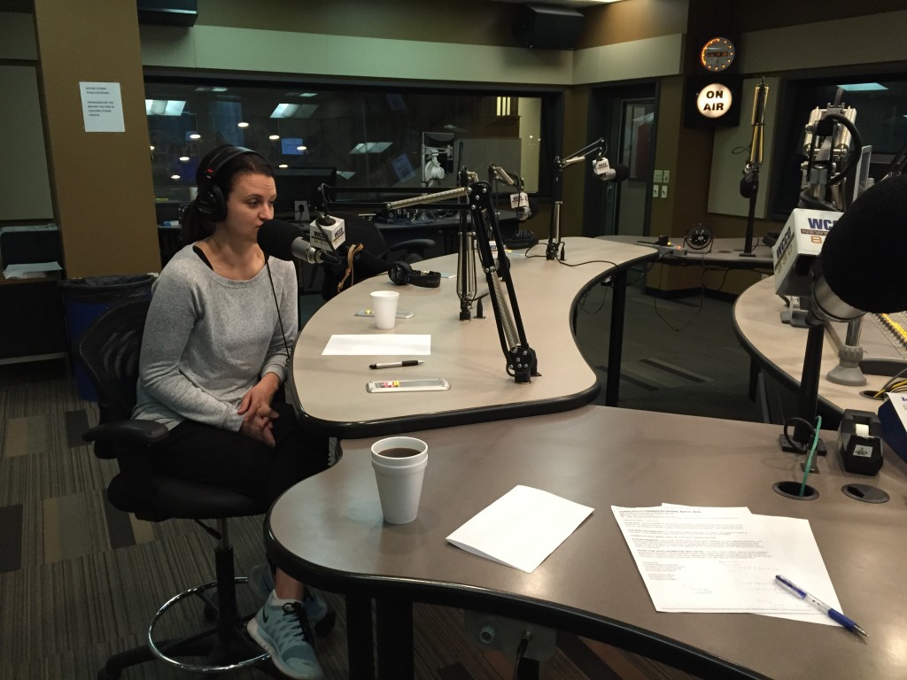
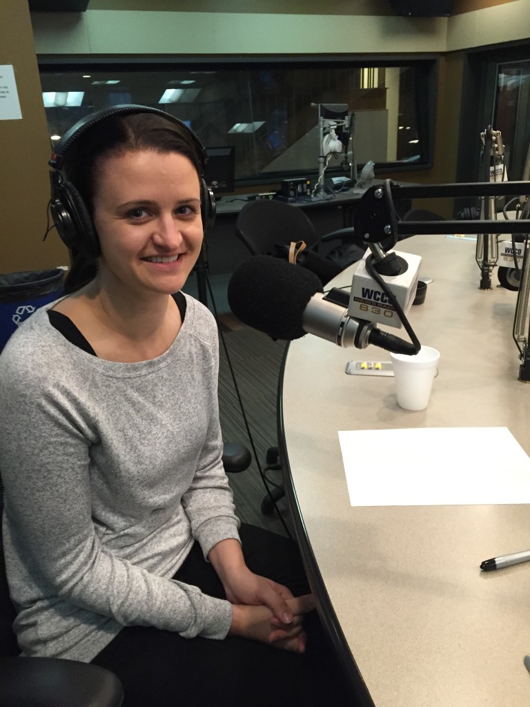
My guest was HCMC ophthalmologist Anne Abel. If you missed the show, be sure to click here to listen to the podcast. She’s a terrific teacher and explains the complexities of the eye really well. That’s her in the picture, giving out serious advice while I distract her by taking pictures!
For instance, here is what we learned today:
- Checking for glaucoma is like pressing your hand on a basketball.
- The lens of the eye is like an M&M.
- Eye doctors drain the fluid from your eye just like a plumber clears a drain.
- The cornea is the windshield of the eye.
That’s all there is to it. Nothing too complicated here. So Dr. Abel is a great surgeon AND she explains things as complex as the eye in a way people can understand. Geez I wish they had taught it that way in medical school!
I’ll touch on a few of the topics we covered on the show today, including M&Ms, basketballs, clogged drains, and windshields – and then I’ll wrap up with some responses to text messages we didn’t get to cover on the broadcast. Read on.
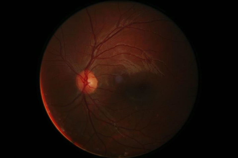
The Mars-like picture above is the back of your eye as seen by your doctor. The bright spot is the optic nerve (literally a portion of your brain visible to the unaided eye) and the lines are blood vessels. Tell me this isn’t cool.
Glaucoma
Dr. Abel’s basketball metaphor is apt. Your eyeball really is like a basketball – only it isn’t filled with air but rather with a gelatinous gooey mass called vitreous humour in the back part and the liquid aqueous humour in the front part. The vitreous comes from the Latin vitrum which means glass and it stays relatively unchanged throughout life. I guess it is shiny and translucent like glass – hence the name. You are born with it and it may serve to keep the structure of the eye intact, but interestingly, an eye doctor can surgically remove it and you will do fine.
The aqueous humour (aqueous coming from the Latin for water) is a different story. This fluid is continuously produced and circulated around the front part of your eye and serves to keep the pressure at the right level. Too much pressure in the eye over time can damage the critical optic nerve which carries visual signals to your brain. Damage the optic nerve and vision loss may result. This is the condition called glaucoma, and it is a leading cause of blindness so it is a big deal. I should mention here, as did Dr. Abel on the radio show, that not all glaucoma fits this neat definition as there are several types and one of them does not feature increased pressure in the eye. So there you go.
For the most part, though, just like pushing on a basketball to check the pressure, your eye doctor is checking the pressure in your eye when testing for glaucoma.
A couple other facts about glaucoma:
- It comes on slowly and is usually painless, so you don’t realize you have it.
- Your peripheral vision (out to the sides) is often affected first, so you may be compensating for that by glancing side-to-side without even knowing it.
- Testing is easy and also painless (they numb the eye surface before testing).
- Treatments exist and they work, but any existing damage to the all-important optic nerve that has already occurred can’t be reversed. Drops can lower eye pressure either by reducing production of the aqueous or by making it easier to drain what fluid is there.
- Laser treatments and surgery are also effective when the drops are not enough – hence the plumber and the drain metaphor (if you don’t know what I’m talking about – listen to the podcast! for more on plumbing and the eye).
For more information on glaucoma, you may want to check out the Glaucoma Research Foundation at glaucoma.org.
Cataract
This is where M&M candy comes in handy. Imagine the lens of your eye, which Dr. Abel tells us is rather like an M&M, presumably not the peanut kind. This structure just inside the opening to your eye helps to focus the image on your retina. Trouble is, as we age the lens gets cloudy, a condition known as cataract. The word itself comes from the Greek for waterfall so I’ve kindly included a picture of a cataract for you. Oops, wrong kind of cataract but it does beg the question about why the eye condition is from the same word as a waterfall. I will leave it to your imagination.
F ortunately, this one is a cinch for an ophthalmologist to fix with a simple surgery. Now it always sounds somewhat disconcerting to me when the procedure is described, but really it is an effective, common, and safe procedure. The lens is simply broken up and removed and a new, synthetic one is implanted.
As we talked about on the air, many people barely even know what they have been missing since cataract, like glaucoma, is slow to develop and is painless. It is easy to diagnose and treat, however, so if your vision isn’t all it used to be and you have reached, ahem, a “certain age” then by all means have a doctor take a peek. You’ll be glad you did.
Cornea – the windshield of the eye
I’m often stymied when trying to describe the cornea – I’m usually fumbling around trying to come up with a good description which ends up something like “the cornea is the clear part overlying the iris, sort of covering up the color part of your eye and it keeps stuff out of the eye” or some such verbiage. On the show today, Dr. Abel was much better at it – the cornea is like the windshield of your eye. Gotta give her credit, that about sums it up without much more elaboration.
Being a clear windshield-like thing, finding a decent picture of just the cornea proved to be too much for my capabilities, so here’s a picture of a regular human eye. Imagine the windshield over those baby blues and that’s the cornea.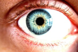
The topic of the cornea came up when a caller asked about pain following a cornea transplant. Too much to get into on this blog post, but cornea transplant (aka keratoplasty) is safe and successful in the vast majority of the cases. It is usually done when your own cornea is damaged due to trauma, infection, previous surgery, and the like and happily the supply (from deceased donors) is usually adequate.
Which brings me to some unanswered texts from the Healthy Matters mailbag. If you are new to the blog . . . I get many texts and phone calls to the live radio broadcast each week – too many to handle on the air. So I try to answer a few every now and then on this blog. Nothing in depth, just a little bit of info for each.
One Healthy Matters listener asked: “What is Fuchs’ dystrophy?”
Pronounced fooks dystrophy, this is a condition in which fluid builds up in your cornea (the windshield) and leads to thickening of the cornea, resulting in blurry vision, sensitivity to light, perhaps a glare overlying the vision. It is more common in women, people over 50, and may be inherited. Treatment usually starts with eyedrops to relieve the swelling but surgery may be needed and is usually quite successful.
Another listener asked if we could “talk about macular degeneration.”
I really wanted to say much more about this condition, as macular degeneration is so common and I don’t get to have an ophthalmologist like Dr. Abel on all that often. She did talk about wet and dry macular degeneration a bit on the air, but since time was short we didn’t get into it in much detail. I think I’ll ask her back for a more in-depth conversation on a future show.
Basically, dry macular degeneration is a thinning of the macula (the part of your retina responsible for seeing straight ahead – your central vision). Compare this to glaucoma in which the peripheral vision usually is affected first. There is no terrific cure for dry macular degeneration, but eye vitamins (like lutein) may help.
The less common wet macular degeneration may come on a bit faster than the slower onset dry type. The wet form is due to overgrowth of blood vessels under the central retina.
Again, macular degeneration is too big a topic for now. Look for a whole radio show on it in the future.
As someone who has a few eye problems of my own – I’ve had surgery at age 3 for lazy eye, I’m basically color-challenged (color-blind, anyone?), and the astigmatism in my left eye means the lens in my eyeglasses on that side is awfully thick – I really appreciate the magnificence of the eye. I hope you’ve learned a bit from the show today and/or this blog post.
 I want to thank Dr. Anne Abel for helping us out on our eye show this morning. To see her or any of my awesome colleagues at HCMC, the number is 612-873-6963 or go to hcmc.org. And as always, please leave me a comment below if you have a question, an idea for a future radio show topic, or you just have something to say!